9 AM, Tesa’s not here yet, but it’s okay – I know what to do now!
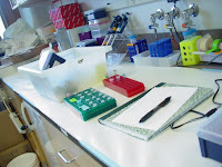
For transfection, I label the microtubes; warm up the media with the water bath; warm up the polymers, DNA, NaAC, Opti-Mem at room temperature; and get different kinds of pipettes, empty tubes, and pipette tips.
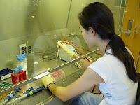
Then I label the cell plates and set up the fume hood.
This is so exciting!!
Transfection is quite a complex process.
Transfection (from the scientific journal I wrote)
One day before a Green Fluorescence Protein (GFP) transfection, a certain type of cell is plated with initial seeding density of 75,000 into two 24-well plates, labeled with all the variables. A plate has 12 wells for 6ug or 3ug of GFP. 6 of the 12 wells, with the same concentration, contain each type of polymers, C32-103, -117, and -122, and liposomes and cells only. Two of the six wells, with the same concentration and polymer type, have the ratio of 20, 30, or 40% polymer to 1% DNA. Two controls are liposome, considered to have relatively low toxicity level, and cells, without gene delivered.
Four hours after the transfection is complete and the gene-delivered cells are put in the incubator, the growth media is changed for all 48 wells and the toxicity levels are reviewed under the microscope by comparing cells-only wells to other wells.
Or simply, different kinds and concentrations of polymers are combined with healthy cells.
Cells are grown, or cultured, in flasks, and when they multiply and fill the bottom of the flasks we need to ‘plate’ them, or divide and transfer them into new flasks. Plating is carried out a day before every transfection.


No comments:
Post a Comment